What is an impacted maxillary canine?
The maxillary (upper) canine, commonly called the “tiger tooth” in Cantonese, plays a very important role in tooth alignment and bite function. It sits between the incisors and the premolars, helps guide the upper and lower jaws smoothly as they bite together, and forms the natural curve of the smile. During growth, however, an upper canine sometimes fails to erupt properly and stays inside the bone or gum. This is called an “impacted maxillary canine”. Impaction is not caused deliberately; during development and eruption, factors such as space, direction or baby teeth prevent the tooth from coming through along its normal path.
1. The basic concept of an impacted maxillary canine
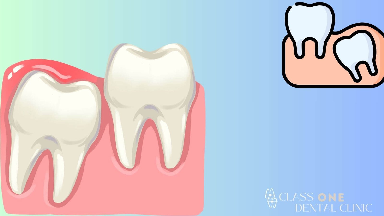
An impacted maxillary canine means that the canine has not appeared in the mouth at the age it should have erupted, while imaging shows that the tooth is present deep in the upper jaw bone or gum but has not broken through the gum or reached the correct position in the dental arch. Such a tooth may be:
1. Completely buried in bone
Both the crown and the root are surrounded by bone, and no sign of the tooth can be seen from the mouth.
2. Partly covered by bone or gum
The crown lies close to the gum surface but shows only a little or none at all, and only a slight bump may be visible.
3. Displaced from the normal dental arch
The canine may erupt, but its direction is too far off, for example on the outer or inner side of the arch, so it does not join the overall alignment.
This kind of impaction mostly involves factors such as insufficient arch space, abnormal growth direction or baby teeth that stay in too long. It does not form in a short time, but develops gradually during childhood and adolescence.
2. Why does an impacted maxillary canine happen?
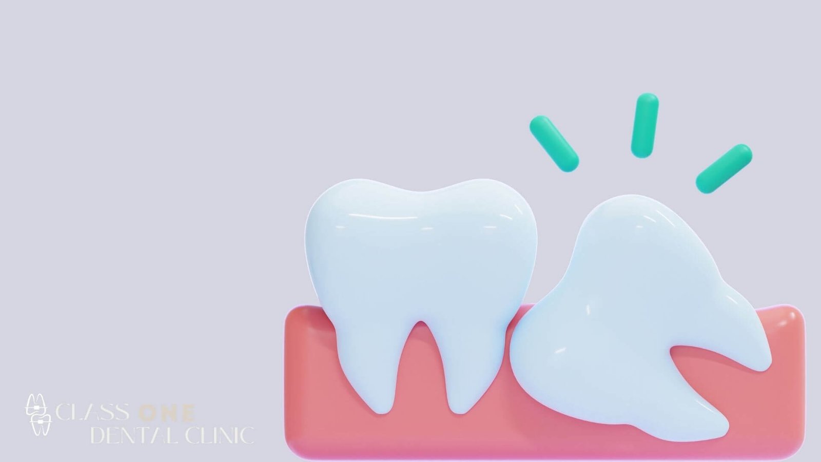
An impacted maxillary canine is usually not caused by a single factor, but by several factors combined. The common causes can be summarised as follows:
(1) Insufficient arch space
The upper canine usually erupts relatively late among the front teeth. If the incisors and other front teeth already occupy most of the space, the arch is not wide enough or the teeth were already crowded, the canine finds it hard to find a suitable position in the existing dentition. Common situations include:
1. A congenitally narrow arch
The upper jaw itself has limited width and the front teeth are already crowded, so the canine is naturally blocked as it comes through.
2. Front teeth that protrude or are crowded
The front teeth are already positioned too far forward or tilted, leaving the dentition “full”. The canine cannot erupt in its normal position and can only move towards the lip side or the palate side, or even stay inside the bone.
(2) Abnormal timing of baby tooth loss
The baby canine should fall out naturally at the right age to make way for the permanent canine. In some cases, however, the baby tooth stays in the arch:
1. The baby canine does not fall out on time
The eruption path of the permanent canine is blocked by the baby tooth, so it can only deviate from the normal direction or stay in the bone.
2. The baby tooth is displaced
The baby tooth has been pushed to one side by external force or alignment problems, making the eruption path of the permanent tooth narrower or distorted.
(3) Abnormal eruption direction
When a tooth develops inside the jaw, if the position of the tooth germ and its eruption direction deviate from the normal course, impaction can also result. Upper canines are especially prone to:
1. A tooth germ that starts too high or close to the floor of the nose
The canine sits too high in the bone, so its eruption route is longer, and if there is also insufficient space it easily stalls.
2. A tilted crown direction
If the canine crown points towards the root of a neighbouring tooth or the bone wall rather than towards the dental arch, it is difficult for it to break through the gum into the correct alignment.
3. Congenital abnormal position
Some cases are related to the position of the tooth germ from birth, and may have some connection with heredity or the pattern of jaw development.
3. What are the effects of an untreated impacted maxillary canine?
An impacted canine is not just a matter of “not seeing that tooth”; it can affect the neighbouring teeth, the bone and the overall bite in several ways.
(1) Pressure on neighbouring teeth
When an impacted canine lies in the bone, its crown or root may come close to, or press against, the roots of neighbouring teeth such as the central or lateral incisors and the premolars. Long-term pressure may lead to:
1. Root resorption of neighbouring teeth
Under continued pressure, the root is gradually “eroded”, weakening its structure or even shortening it.
2. Neighbouring teeth becoming loose or shifting position
Once the root is damaged, the stability of the tooth falls, and it may become loose or move, affecting appearance and bite.
(2) Formation of cysts or other lesions
If the tissue around an impacted tooth, such as the dental follicle, develops disease under certain conditions, a cyst or other pathological change may form. Such a cyst can:
1. Expand and destroy bone
The cyst gradually grows and erodes the jaw bone, thinning the local bone or leaving a defect.
2. Affect neighbouring teeth and structures
The surrounding roots may be involved, and the teeth may become sensitive or loose, or need further treatment.
(3) Effects on the bite and function
The canine has a guiding role in the bite, helping the upper and lower jaws glide smoothly in side-to-side movements and protecting the molars from excessive sideways pressure. If an upper canine is impacted and does not take part in the bite, this may cause:
1. An unbalanced bite
With one important tooth missing, chewing forces are distributed unevenly in some directions, which over time can increase the load on other teeth.
2. A change in wear patterns
Molars or other teeth may take extra pressure from standing in for the canine’s bite-guiding role, and show abnormal wear.
3. Effects on the appearance of the whole dentition
The canine has a clear presence in the smile, and when it is impacted or missing the curve of the arch may look unnatural, affecting confidence in appearance.
4. Common approaches to treating an impacted maxillary canine
The specific treatment plan must be drawn up by a dentist after a comprehensive examination, imaging analysis and bite assessment. The following only outlines common approaches, for conceptual understanding.
(1) Monitoring and space management (childhood and adolescence)
During the growth period, if the canine’s eruption path shows a tendency towards impaction, the dentist may:
1. Assess when the baby canine falls out
If the baby tooth has stayed in too long, timely extraction may be recommended so that the permanent canine has room.
2. Carry out space management
If the arch is markedly crowded, orthodontic treatment (braces) may be considered to expand the space or redistribute the tooth positions, leaving room for the canine.
(2) Surgical exposure combined with orthodontic traction
When the canine is already impacted but its position is still usable, the common approach is:
1. Surgical exposure of the crown
A specialist performs a minor operation to expose the part of the crown buried in the gum or bone.
2. Traction of the tooth with an orthodontic appliance
An orthodontic attachment is bonded to the canine crown, and the pull of the braces gradually guides the canine into its correct position in the arch.
This approach takes longer and requires good oral hygiene, but it preserves the natural canine, balancing function and appearance.
(3) Extraction of the impacted canine and alternatives
If the impacted canine is in a very unfavourable position, is causing clear damage to neighbouring teeth and bone, or has a poor prognosis itself, the dentist may assess the case and recommend extraction. After extraction, the following need to be considered:
1. Keeping or adjusting the space in the dentition
Orthodontic treatment or other methods can be used to adjust the dentition and reduce the impact of the missing tooth on appearance and bite.
2. Future restoration
Depending on the situation, dentures, a dental implant or other restorative methods may be considered to restore function and appearance.
5. When should you suspect an impacted maxillary canine?
In general, the permanent canines should erupt progressively during adolescence. If any of the following occurs, it is advisable to consult a dentist as soon as possible:
1. At the expected age, the canine on one or both sides has still not appeared
The other front teeth erupted long ago, but there is still no sign of the canine.
2. A persistent bump on the gum, or an unusual protrusion
Especially at the front of the upper jaw near the side of the nose or higher on the gum, with a hard lump feeling.
3. Neighbouring teeth become unexplainably loose or move
For example, an incisor shifts for no clear reason, or imaging shows an abnormal root condition.
In these situations, having the dentist arrange imaging (such as X-rays or 3D imaging) to confirm the position of the canine and the surrounding structures is the key first step in diagnosis and management.
6. Impacted maxillary canines and self-care
Even though the impaction itself must be treated by medical means, patients can still pay attention to the following in daily life:
1. Get children into the habit of regular dental check-ups early
Regular check-ups during the mixed dentition stage allow the dentist to spot abnormal baby tooth loss timing or arch space problems early.
2. Maintain good oral hygiene
If surgery and orthodontic traction are needed, oral hygiene is even more important, to reduce the risk of inflammation and infection.
3. Do not guess or delay
If an impacted tooth is suspected, seek professional advice early rather than waiting for years, so that any lesion does not enlarge or affect neighbouring teeth.
Identifying and managing impacted teeth involves imaging, diagnosis and multidisciplinary cooperation, and cannot be known from appearance alone. Understanding the concept helps patients be clearer about their own condition and the direction of treatment when they receive a related diagnosis.
Impacted maxillary canine FAQ
Question 1: Does an impacted maxillary canine always require tooth extraction?
Not all impacted canines have to be removed. Whether extraction is needed depends on the position of the canine, its relationship with neighbouring teeth, whether it is causing root resorption or a cyst, and whether it can be brought back into the arch by orthodontic traction. In some cases, surgical exposure combined with braces can preserve the canine and restore normal alignment; in others, extraction is needed because of its position and pathological factors.
Question 2: If an impacted maxillary canine is only discovered in adulthood, is treatment more difficult?
In adulthood the jaw bone is more stable and teeth move relatively more slowly, so traction of an impacted canine may be more difficult. But how difficult it is is not decided by age alone; it depends on the position of the impacted tooth, the shape of the root and the condition of the surrounding bone. Adult patients can still discuss feasible options in detail with their dentist and choose according to the risks and expected results.
Question 3: If there is no discomfort, can an impacted canine simply be ignored?
Even if there is no obvious pain or functional problem for now, an impacted canine still carries potential risks, such as pressure on the roots of neighbouring teeth, cyst formation or an unbalanced bite later on. It is generally recommended that a dentist at least carry out a comprehensive assessment to understand its position and the surrounding tissues, and then decide between observation, orthodontic treatment or another approach, rather than ignoring it completely.